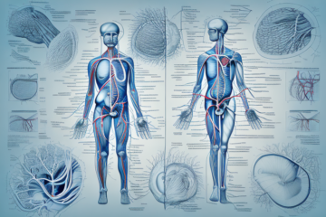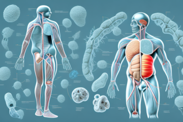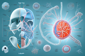
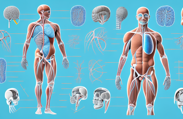
The urinary system is an incredible network of organs that work together to remove waste products from the body and maintain a healthy balance of fluids, electrolytes, and minerals. Ureters, in particular, play a vital role in this system by providing a pathway for urine to travel from the kidneys to the bladder. In this article, we’ll be discussing the various aspects of the ureters, including their function, anatomy, diseases, and treatments.
The Role of Ureters in the Urinary System
The ureters are two narrow tubes, each about 10-12 inches long, that are responsible for transporting urine from the kidneys to the bladder. They are lined with smooth muscle that contracts and relaxes in a rhythmic manner, pushing the urine towards the bladder through a process called peristalsis. The ureters play a crucial role in maintaining the balance of fluids and electrolytes in the body by helping to excrete waste products from the body efficiently.
In addition to their role in transporting urine, the ureters also have a protective function. The muscular walls of the ureters help to prevent urine from flowing back up into the kidneys, which could cause damage or infection. The ureters also have a valve-like mechanism at the point where they enter the bladder, which helps to prevent urine from flowing back up into the ureters. This ensures that urine flows in one direction, from the kidneys to the bladder, and helps to maintain the health of the urinary system.
How Ureters Transport Urine from the Kidneys to the Bladder
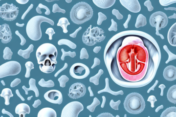
The process of urine transport from the kidneys to the bladder starts in the renal pelvis, which is the central collecting area of each kidney. The urine is then passed into the ureters, which run down behind the abdominal cavity and terminate at the bladder. The opening of the ureters into the bladder is guarded by a one-way valve, or sphincter, which prevents the urine from backing up into the kidneys and causing damage.
The ureters are muscular tubes that contract rhythmically to move urine from the kidneys to the bladder. This peristaltic movement is similar to the way the esophagus moves food down to the stomach. The ureters also have a lining of smooth muscle that helps to push the urine along.
In some cases, a blockage can occur in the ureters, preventing urine from flowing properly. This can be caused by a variety of factors, including kidney stones, tumors, or scar tissue. If left untreated, a blockage can lead to kidney damage or infection. Treatment options for a blocked ureter may include surgery, medication, or the use of a stent to keep the ureter open.
The Structure and Layers of the Ureters
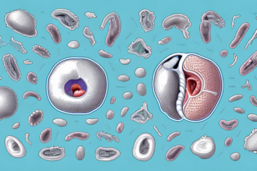
The walls of the ureters are made up of three distinct layers. The innermost layer, also known as the mucosa, is made up of transitional epithelial cells that allow the ureters to expand and contract as urine passes through. The middle layer is composed of smooth muscle, which contracts to push the urine along. The outermost layer, or adventitia, is made up of connective tissues and blood vessels that supply the ureters with oxygenated blood.
The ureters are tubes that connect the kidneys to the bladder and play a crucial role in the urinary system. The ureters are approximately 10-12 inches long and are responsible for transporting urine from the kidneys to the bladder. The walls of the ureters are lined with smooth muscles that contract rhythmically to move urine along the tubes. The ureters also have valves that prevent urine from flowing back into the kidneys, ensuring that urine flows in one direction only.
Common Disorders and Diseases of the Ureters
Several diseases and disorders can affect the ureters, and some of the most common ones are ureteral obstruction, ureteral reflux, and kidney stones. Ureteral obstruction occurs when there is a blockage in one or both of the ureters, which can cause urine to back up and lead to complications like kidney damage and infection. Ureteral reflux is a condition where urine flows back into the ureters from the bladder, which can cause inflammation and infection. Kidney stones, on the other hand, are hard masses of minerals that can form in the kidneys and travel down the ureters towards the bladder.
Other less common disorders of the ureters include ureteral strictures, which are narrowings of the ureters that can cause urine to back up and lead to kidney damage, and ureteral tumors, which can be cancerous or non-cancerous growths that can block the flow of urine. Additionally, some people may be born with abnormalities in their ureters, such as a duplicated ureter or a ureterocele, which can cause urinary tract infections and other complications.
Diagnosis and Treatment Options for Ureteral Obstruction
Diagnosis of ureteral obstruction often involves imaging tests like a CT scan or ultrasound, as well as urine tests and blood tests to check for signs of infection or kidney damage. Treatment for ureteral obstruction may depend on the underlying cause and severity of the condition, but options include medications, surgery, or minimally invasive procedures like stenting or laser lithotripsy.
In some cases, ureteral obstruction may be caused by a tumor or other abnormal growth in the urinary tract. In these cases, treatment may involve a combination of surgery and radiation or chemotherapy to remove the growth and prevent it from returning.
For patients with chronic or recurrent ureteral obstruction, ongoing monitoring and management may be necessary to prevent complications like kidney damage or infection. This may involve regular imaging tests and check-ups with a urologist or other specialist to ensure that the condition is being properly managed and treated.
Understanding Ureteral Reflux and Its Causes
Ureteral reflux is usually diagnosed through a voiding cystourethrogram test, which involves the use of X-rays to track the flow of urine from the bladder back towards the ureters. Treatment options may include medications or bladder training exercises to help improve bladder function and prevent reflux from occurring.
It is important to note that ureteral reflux can lead to serious complications if left untreated. These complications may include kidney damage, urinary tract infections, and even kidney failure in severe cases. Therefore, it is crucial to seek medical attention if you suspect you may have ureteral reflux.
In some cases, surgery may be necessary to correct ureteral reflux. This may involve repositioning the ureters or using a surgical procedure to create a valve-like mechanism to prevent urine from flowing back into the ureters. Your doctor will determine the best course of treatment based on the severity of your condition and your overall health.
The Relationship Between Kidney Stones and Ureter Pain
When a kidney stone gets stuck in the ureter, it can cause excruciating pain that radiates from the back into the abdomen and groin. Treatment options for kidney stones typically include pain management, the use of medications to help pass the stone naturally, or in some cases, surgery may be needed to remove the stone.
It is important to note that certain factors can increase the risk of developing kidney stones, such as a diet high in salt and animal protein, dehydration, and a family history of kidney stones. Additionally, some medical conditions, such as hyperparathyroidism and inflammatory bowel disease, can also increase the risk of kidney stone formation.
If left untreated, kidney stones can lead to complications such as urinary tract infections, kidney damage, and even sepsis. It is important to seek medical attention if you experience symptoms of a kidney stone, such as severe pain, nausea, vomiting, and blood in the urine.
Preventing Infections of the Ureters Through Lifestyle Changes
Preventing infections of the ureters involves making simple lifestyle changes, such as drinking plenty of water, practicing good hygiene, avoiding irritants like bubble baths or scented products, and seeking prompt treatment for urinary tract infections (UTIs).
Another important lifestyle change to prevent infections of the ureters is to urinate frequently and completely. Holding in urine for long periods of time can increase the risk of UTIs and other urinary tract infections. Additionally, wiping from front to back after using the bathroom can help prevent the spread of bacteria from the anus to the urethra.
In some cases, individuals may be more prone to ureter infections due to underlying medical conditions or anatomical abnormalities. In these cases, it is important to work closely with a healthcare provider to develop a personalized prevention plan that may include medication or other interventions.
Investigating Lower Back Pain: Could it be Related to Your Ureters?
Lower back pain can be a sign of ureteral obstruction or kidney stones, especially if the pain is severe and comes on suddenly. It’s important to seek medical attention if you experience persistent or severe lower back pain, as it could be a sign of a more serious underlying condition.
Ureters are tubes that carry urine from the kidneys to the bladder. When these tubes become obstructed, urine can back up into the kidneys, causing pain and discomfort in the lower back. This condition is known as hydronephrosis and can lead to kidney damage if left untreated.
Other symptoms of ureteral obstruction or kidney stones may include pain or discomfort during urination, blood in the urine, and nausea or vomiting. Treatment options may include medication, surgery, or shock wave lithotripsy, a non-invasive procedure that uses sound waves to break up kidney stones.
Surgical Techniques for Repairing or Replacing Damaged Ureters
In some cases, surgical techniques may be required to repair or replace damaged ureters. These procedures may include re-implantation of the ureters, surgical reconstruction, or the use of tissue grafts to replace damaged sections of the ureters.
Re-implantation of the ureters involves detaching the ureter from the bladder or kidney and reattaching it in a new location. This procedure is often used to treat ureteral strictures, which are narrow sections of the ureter that can cause urine to back up into the kidney.
Surgical reconstruction of the ureter may be necessary in cases where the ureter has been severely damaged or blocked. This procedure involves removing the damaged section of the ureter and reconnecting the remaining healthy sections. In some cases, a stent may be placed to keep the ureter open during the healing process.
How Imaging Tests Help Detect Abnormalities in the Ureters
Imaging tests like CT scans, MRIs, or ultrasounds are often used to detect abnormalities in the structure or function of the ureters. These tests can help doctors diagnose conditions like ureteral obstruction, ureteral reflux, or kidney stones, and develop an appropriate treatment plan.
In conclusion, understanding the role and function of the ureters is essential for maintaining good urinary health. By making the necessary lifestyle changes, seeking prompt medical attention when needed, and following prescribed treatments, individuals can ensure that their ureters function efficiently and effectively in removing waste products from the body.
It is important to note that imaging tests are not always necessary for detecting abnormalities in the ureters. In some cases, a simple urine test or physical examination may be enough to identify a problem. However, imaging tests can provide a more detailed and accurate picture of the ureters, which can be especially helpful in cases where the diagnosis is unclear or the condition is more complex.